Sketch And Label Of A Cross Section Of A Long Bone - Long Bone Cross Section Worksheet Teaching Resources. Sketch and label a cross section of a bone. You need to get 100% to score the 10 points available. Bone remodeling and repair 11. This is an online quiz called long bone anatomy. Once we stop growing (between 18.
On this page, you will find two images i created that illustrate the parts of a long bone and long bone structure. Bone remodeling and repair 11. Osteons are oriented parallel to the diaphysis of the long bone. This is the long central shaft. Sketch and label of a cross section of a long bone.
Long Bone Wikipedia from upload.wikimedia.org Once we stop growing (between 18. Related posts of cross section of human bone diagram foot bone anatomy x ray. Label the haversian canal, osteocyte (mature bone cell) in lacuna, and canaliculi. Draw and label the following structures as they appear using the 10x objective o bone marrow o bony trabeculae activity 5.2.3: Make sure learners follow all the criteria for a biological drawing. Draw and label a longitudinal section of a long bone. Plates of cartilage, also known as growth plates which allow the long bones to grow during childhood. The osteocytes are arranged in concentric rings of bone matrix called lamellae (little plates), and their processes run in interconnecting canaliculi.
The digital cushion sits just behind the pedal bone and above the sensitive frog.
This is an online quiz called long bone anatomy. A typical long bone shows the gross anatomical characteristics of bone. You need to get 100% to score the 10 points available. Related posts of cross section of human bone diagram foot bone anatomy x ray. Once we stop growing (between 18. Forms the larger rounded ends of long bones. This is the long central shaft. A long bone has two parts: Then, fill in the table below to describe each. At the elbow, it connects primarily to the ulna, as the forearm's radial bone connects to the. Bone matrix and cells bone matrix osseous tissue is a connective tissue and like all connective tissues contains relatively few cells and large amounts of extracellular matrix. Long bones have a thick outside layer of compact bone and an inner medullary cavity containing bone marrow. The structure of a long bone consists of several sections:.
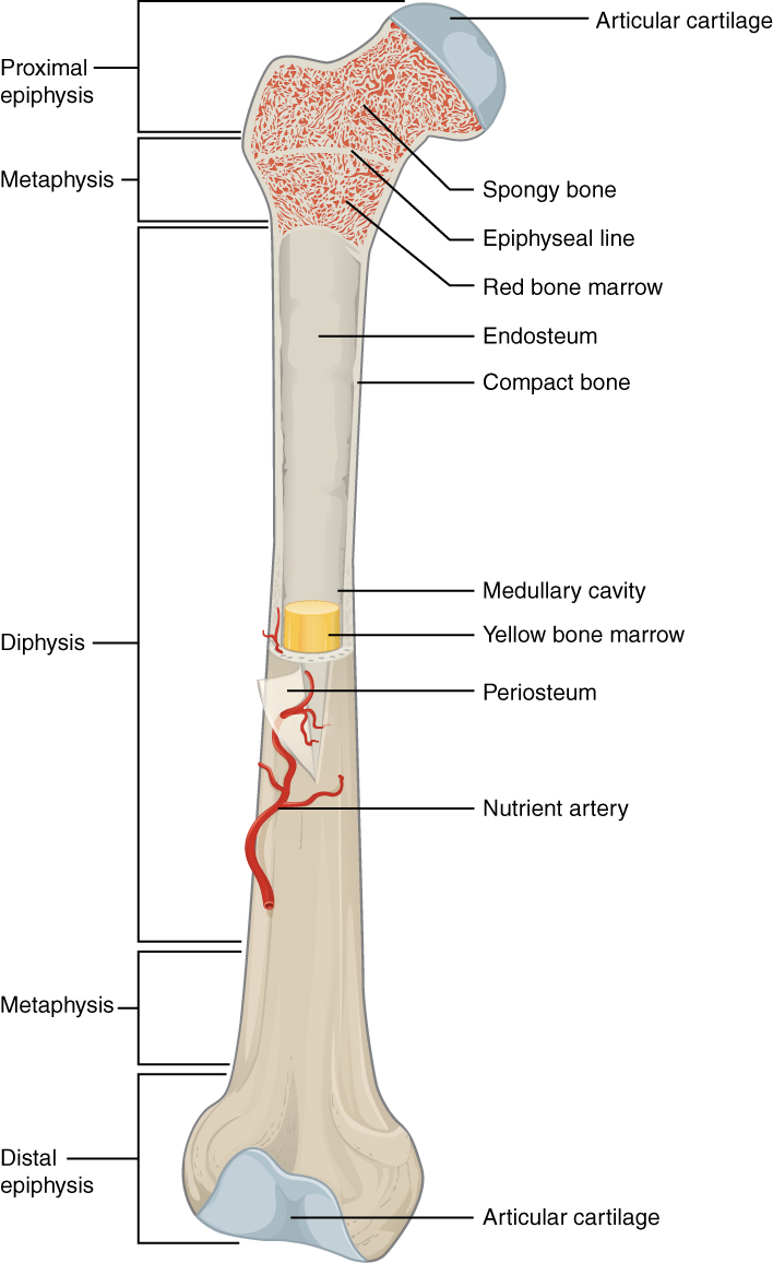
The periosteum contains many strong collagen fibers that are used to firmly anchor tendons and muscles to the bone for movement. In the space provided draw a longitudinal section of a long bone and label the following parte proximal epiphysis, distal epiphysis, diaphysis, metaphysis, medullary cavity, epiphyseal line 2. Cross section of long bone. The outside of a bone is covered in a thin layer of dense irregular connective tissue called the periosteum. A long bone is a bone that has greater length than width.
Bone Structure Anatomy And Physiology I from s3-us-west-2.amazonaws.com This is an online quiz called label the long bone. Use colored pencils to draw and label the following structures as they appear using the 40x objective, or by looking at an image from the internet. The following slides will help show the several methods or types of section views Also known as the middle phalanx, the short pastern bone sits on top of the articulating joint of the pedal bone and underneath the long pastern bone. It is located between the elbow joint and the shoulder. The diaphysis is the tubular shaft that runs between the proximal and distal ends of the bone. A long bone has two parts: Continue to label this drawing as you explore the inside of the bone.
Anatomy of a long bone 1.
Bone matrix and cells bone matrix osseous tissue is a connective tissue and like all connective tissues contains relatively few cells and large amounts of extracellular matrix. Area between the diaphysis and epiphysis at both ends of the bone. The diaphysis is the tubular shaft that runs between the proximal and distal ends of the bone. Label the haversian canal, osteocyte (mature bone cell) in lacuna, and canaliculi. Erythrocytes, or red blood cells, are by far the predominant cell type in the blood smear. The humerus is the long bone in the upper arm. (do not copy and paste a picture from the text or internet.) The ends of a long bone contain spongy bone and an epiphyseal line. The diaphysis and the epiphysis. The head of each end of a long bone consists largely of spongy bone and is covered with hyaline cartilage. The diaphysis and the epiphysis. Bone remodeling and repair 11. This is an online quiz called label the long bone.
The humerus is the long bone in the upper arm. Plates of cartilage, also known as growth plates which allow the long bones to grow during childhood. The osteocytes are arranged in concentric rings of bone matrix called lamellae (little plates), and their processes run in interconnecting canaliculi. There is a printable worksheet available for download here so you can take the quiz with pen and paper. Smartdraw includes 1000s of professional healthcare and anatomy chart templates that you can modify and make your own.
Histology Laboratory Manual from www.columbia.edu Cross section of a long bone. The diaphysis and the epiphysis. The ends of a long bone contain spongy bone and an epiphyseal line. Anatomy of a long bone 1. External circumferential lamellae, osteon, central canal, perforating canals, lacuna, canaliculi, concentric lamellae. Draw and label the following structures as they appear using the 10x objective o bone marrow o bony trabeculae activity 5.2.3: Click on the tags below to find other quizzes on the same subject. End of a long bone.
The diaphysis and the epiphysis.
A section view is a view used on a drawing to show an area or hidden part of an object by cutting away or removing some of that object. It suggests that the bone will have equal strength in all directions. Click on the tags below to find other quizzes on the same subject. Create a drawing of the bone section in your laboratory journal and label the areas listed above. Area between the diaphysis and epiphysis at both ends of the bone. The outside of a bone is covered in a thin layer of dense irregular connective tissue called the periosteum. Plates of cartilage, also known as growth plates which allow the long bones to grow during childhood. Terms in this set (3) epiphysis. The next section will discuss the identification of the immature cells of the bone marrow. You need to get 100% to score the 10 points available. Cow and human long bones have a similar general structure. The end of a growing tibia, cut lengthwise*. Looking at a bone in cross section, there are several distinct layered regions that make up a bone.
Share this post
0 Response to "Sketch And Label Of A Cross Section Of A Long Bone - Long Bone Cross Section Worksheet Teaching Resources"


0 Response to "Sketch And Label Of A Cross Section Of A Long Bone - Long Bone Cross Section Worksheet Teaching Resources"
Post a Comment